Proteomic characteristics of resistant Proteus species in women with urinary tract infections in Nasarawa State, Nigeria

Authors

  • M. H. Muhammad
    Department of Microbiology, University Teaching Hospital, Lafia, Nigeria.
  • O. O. Orole
    Department of Microbiology, Federal University of Lafia, Nasarawa State, Nigeria.
  • J. F. Nfongeh
    Department of Microbiology, Federal University of Lafia, Nasarawa State, Nigeria.
  • E. S. Audu
    Department of Medical Microbiology, Federal University of Lafia, Nasarawa State, Nigeria.
  • F. Gbadeyan
    Department of Microbiology, Federal University of Lafia, Nasarawa State, Nigeria.

Keywords:

Multidrug resistance, Virulence, Plasmids, Outer membrane proteins, Bacteriuria

Abstract

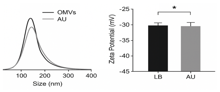
Urinary tract infections (UTIs) represent a recurring clinical challenge, primarily due to escalating pathogen resistance. Although Proteus species are not the most commonly isolated pathogens in UTIs, they typically exhibit high levels of resistance when present. This study investigated the proteomic profiles of resistance and virulence determinants in Proteus species isolated from women with UTIs in Nasarawa State, Nigeria. Urine samples were collected from 368 patients, from which Proteus isolates were identified and evaluated for resistance and virulence factors. Proteomic characteristics were determined following growth in Luria–Bertani (LB) broth and artificial urine (AU). Ten Proteus isolates, comprising four P. vulgaris and six P. mirabilis, were obtained from 21 urine samples exhibiting significant bacteriuria. Both species were found to harbor adhesion (20 kb) and hemolysin (30 kb and 50 kb) plasmids. Identified resistance elements included an ESBL plasmid (40 kb and 80 kb), a conjugative R-plasmid (50 kb), and a multidrug conjugative plasmid (200 kb). The outer membrane vesicle (OMV) yield ranged from 89.5-137μg/mL for P. vulgaris and 97.6-140μg/mL for P. mirabilis. The OMVs exhibited a unimodal size distribution, with average diameters ranging from 70 nm to 180 nm. Recovered outer membrane-associated proteins included outer membrane protein A (OmpA), porins (OmpC and OmpF), flagellin structural proteins (FliC and FlgE), lipoproteins (Lpp and Pal), and proteases (DegP and HtrA). Notably, P. mirabilis cultured in AU was enriched with virulence-related proteins, such as urease subunits (UreA, UreB, and UreC) and fimbrial adhesion proteins. This study confirms the complex nature of virulence and resistance factors in Proteus species, which significantly contribute to treatment failure in patients. 0.45μm

Dimensions

[1] A. Flores-Mireles, J. N. Walker, M. Caparon & S. J. Hultgren “Urinary tract infections: epidemiology, mechanisms of infection and treatment options”, Nat Rev Microbiol. 13 (2015) 269. https://doi.org/10.1038/nrmicro3432.

[2] K. Czajkowski, M. Broś-Konopielko & J. Teliga-Czajkowska, “Urinary tract infection in women”, Przegl Menopauzalny 20 (2021) 1. https://doi.org/10.5114/pm.2021.105382.

[3] J. Esther, O. B. Onyebuchi, O. E. Eugenia C. A. Ogbonna, M. A. Nwaodu, I. G. Chiaka, O. Folaranmi, S. O. Nduka, N. Folaranmi & I. B. Umeh, “Antimicrobial resistance in Nigeria’s healthcare system: a comprehensive narrative review and policy implications”, Discov. Public Health 22 (2025) 460. https://doi.org/10.1186/s12982-025-00859-1.

[4] N. A. Hassuna, D. N. Kotb, M. Lami & S. S. Abdelrahim, “Characterization of antimicrobial resistance among proteus mirabilis isolates from catheter-associated urinary tract infections and non-catheter-associated urinary tract infections in Egypt”, BMC Infectious Diseases 25 (2025) 767. https://doi.org/10.1186/s12879-025-11118-8.

[5] O. O. Orole & N. S. Hadi, “Characterization and plasmid profile of resistant klebsiella pneumoniae isolates in patients with urinary tract infection in Nasarawa State, Nigeria”, Int. J. Appl. Sci. Biotechnol 8 (2020) 21. https://doi.org/10.3126/ijasbt.v8i1.28252.

[6] E. C. Mantu, J. F. Nfongeh, O. O. Orole, S. Obiekezie & J. N. Lamini, “Bacterial pathogens associated with urinary tract infections among rural women in Doma, Nasarawa State, Nigeria”, Int. J. Curr. Microbiol. App Sci. 7 (2018) 2823. https://doi.org/10.20546/ijcmas.2018.708.297.

[7] L. Naing, T. Winn & B. N. Rusli. “Practical issues in calculating the sample size for prevalence studies”, Arch. Orofac Sci. 1 (2006) 9. https://api.semanticscholar.org/CorpusID:961076.

[8] U. Nwankwo, E. Chinenye, E. Joachim & O. Ikechukwu, “Prevalence of uropathogens and associated risk factors in urinary tract infection in pregnant women attending antenatal clinic in a teaching hospital in Awka, Nigeria”, J. Adv. Microbiol. 24 (2024) 104. https://doi.org/10.9734/jamb/2024/v24i4819.

[9] A. Bhugra & S. Gachinmath, “Significant bacteriuria among requested repeat urine samples and its clinical correlation”, Iran J. Microbiol. 13 (2021) 592. https://doi.org/10.18502/ijm.v13i5.7421.

[10] E. A. Gumar, A. S. Hamzah & W. F. Hamad, “Study of some resistance genes in clinical proteus mirabilis”, Archives of Razi Institute 6 (2022) 2235. https://doi.org/10.22092/ARI.2022.358489.2230.

[11] B. D’Alessandro, L. M. Lery, W. M. Von-Krüger, A. Lima, C. Piccini & P. Zunino, “Proteomic analysis of proteusmirabilis outer membrane proteins reveals differential expression in vivo vs. in vitro conditions”, FEMS Immunol. Med. Microbiol. 13 (2011) 174. https://doi.org/10.1111/j.1574-695X.2011.00839.x.

[12] V. Stenbakk, M. Jakubec, T. Vasskog, T. Kristoffersen, J. P. Cavanagh, J. U. Ericson, J. Isaksson & G. E. Flaten, “Bacterial extracellular vesicles: towards realistic models for bacterial membranes in molecular interaction studies by surface plasmon resonance”, Front Mol. Biosci. 10 (2023) 1277963. https://doi.org/10.3389/fmolb.2023.1277963.

[13] B. Baimakhanova, A. Sadanov, L. Trenozhnikova & et al, “Understanding the burden and management of urinary tract infections in women”, Diseases (Basel) 13 (2025) 59. https://doi.org/10.3390/diseases13020059.

[14] O. Storme, J. T. Saucedo, A. Garcia-Mora, M. Dehesa-Dávila & K. G. Naber, “Risk factors and predisposing conditions for urinary tract infection”, Therapeutic Advances in Urology 11 (2019) 1756287218814382. https://doi.org/10.1177/1756287218814382.

[15] M. H. Muhammad, J. F. Nfongeh, S. L. Ageba & O. O. Orole, “Determinants of virulence and antimicrobial resistance in proteus species from women with urinary tract infections in Lafia, Nigeria”, Innov. Med. Omics (2025) 025500073. https://doi.org/10.36922/IMO025500073.

[16] G. Mancuso, A. Midiri, E. Gerace, M. Marra, S. Zummo & C. Biondo, “Urinary tract infections: the current scenario and future prospects”, Pathogens 12 (2023) 623. https://doi.org/10.3390/pathogens12040623.

[17] H. S. Yaseen, Q. N. A. Thewaini & Z. M. Jassim, “Prevalence and role of adhesion genes in proteus mirabilis”, SAR J. Pathol Microbiol 5 (2024) 148. https://doi.org/10.36346/sarjpm.2024.v05i04.007.

[18] M. Zhuang, W. Yan, Y. Xiong & et al, “Horizontal plasmid transfer promotes antibiotic resistance in selected bacteria in Chinese frog farms”, Environ. Int. 190 (2024) 108905. https://doi.org/10.1016/j.envint.2024.108905.

[19] S. Castaneda-Barba, E. M. Top & T. Stalder, “Plasmids, a molecular cornerstone of antimicrobial resistance in the One Health era”, Nat. Rev. Microbiol. 22 (2023) 18. https://doi.org/10.1038/s41579-023-00926-x.

[20] M. J. González, N. Navarro, E. Cruz, S. Sánchez, J. O. Morales, P. Zunino, L. Robino, A. Lima & P Scavone, “First report on the physicochemical and proteomic characterization of Proteus mirabilis outer membrane vesicles under urine-mimicking growth conditions: comparative analysis with Escherichia coli”, Frontiers in Microbiology 15 (2024) 1493859. https://doi.org/10.3389/fmicb.2024.1493859.

[21] H. Hariharan, Y. Kesavan, H. S. Mohideen, N. S. Raja & S. Loganathan, “The composition of the bacterial culture media influences the secretion and sorting of proteins into the bacterial extracellular vesicle”, J. Pure Appl. Microbiol. 20 (2026) 345. https://doi.org/10.22207/JPAM.20.1.21.

[22] C. Schwechheimer & M. J. Kuehn, “Outer-membrane vesicles from gram-negative bacteria: biogenesis and functions”, Nat. Rev. Microbiol. 13 (2015) 605. https://doi.org/10.1038/nrmicro3525.

[23] D. Szczerbiec, S. Glińska, J. Kamińska & D. Drzewiecka, “Outer membrane vesicles formed by clinical proteusmirabilis strains may be incorporated into the outer membrane of other P. mirabilis Cells and demonstrate lytic properties”, Molecules 29 (2024) 4836. https://doi.org/10.3390/molecules29204836.

[24] M. J. González, N. Navarro, E. Cruz, S. Sánchez, J. O. Morales, P. Zunino, L. Robino, A. Lima & P. Scavone, “First report on the physicochemical and proteomic characterization of Proteus mirabilis outer membrane vesicles under urine-mimicking growth conditions: comparative analysis with Escherichia”, Coli. Front Microbiol. 15 (2024) 1493859. https://doi.org/10.3389/fmicb.2024.1493859.

[25] L. Denzer, H. Schroten & C. Schwerk, “From gene to protein—How bacterial virulence factors manipulate host gene expression during infection”, Int. J. Mol. Sci. 21 (2020) 3730. https://doi.org/10.3390/ijms21103730.

[26] G. Midekessa, K. Godakumara, J. Ord, J. Viil, F. Lättekivi, K. Dissanayake, S. Kopanchuk, A. Rinken, A. Andronowska, S. Bhattacharjee, T. Rinken & A. Fazeli, “Zeta potential of extracellular vesicles: toward understanding the attributes that determine colloidal stability”, ACS Omega. 5 (2020) 16701. https://doi.org/10.1021/acsomega.0c01582.

[27] Y. M. Gnopo, A. Misra, H. L. Hsu, M. P. DeLisa, S. Daniel & D. Putnam, “Induced fusion and aggregation of bacterial outer membrane vesicles: experimental and theoretical analysis”, J. Colloid Interface Sci. 758 (2020) 068. https://doi.org/10.1016/j.jcis.2020.04.068.

[28] B. D. Needham & M. S. Trent, “Fortifying the barrier: the impact of lipid a remodeling on bacterial pathogenesis”, Nat. Rev. Microbiol. 11 (2013) 467. https://doi.org/10.1038/nrmicro3047.

[29] E. J. O’Donoghue & A. M. Krachler, “Mechanisms of outer membrane vesicle entry into host cells”, Cell Microbiol. 18 (2016) 1508. https://doi.org/10.1111/cmi.12655.

[30] M. Toyofuku, N. Nomura & L. Eberl, “Types and origins of bacterial membrane vesicles”, Nature Reviews Microbiology 17 (2019) 13. https://doi.org/10.1038/s41579-018-0112-2.

[31] N. Kaderabkova, M. Bharathwaj, R. C. D. Furniss, D. Gonzalez, T. Palmer & D. A. Mavridou, “The biogenesis of $beta$-lactamase enzymes”, Microbiology 168 (2022) 001217. https://doi.org/10.1099/mic.0.001217.

[32] J. Wang, Q. Liu, F. Huang, S. Wang, C. Wu, Y. Chao, S. Huang, Y. Liang & X. Pan, “Bacterial outer membrane vesicles: a smart platform for biomedical applications”, Cell Commun. Signal 7 (2026) 24. https://doi.org/10.1186/s12964-025-02645-7.

[33] A. T. Jan, “Outer membrane vesicles (OMVs) of gram-negative bacteria: a perspective update”, Front Microbiol. 8 (2017) 1053. https://doi.org/10.3389/fmicb.2017.01053.

Average zeta potential comparison of OMV from both media

Published

2026-05-20

How to Cite

Proteomic characteristics of resistant Proteus species in women with urinary tract infections in Nasarawa State, Nigeria. (2026). African Scientific Reports, 5(2), 456. https://doi.org/10.46481/asr.2026.5.2.456

Issue

Section

BIOLOGICAL SCIENCES SECTION

How to Cite

Proteomic characteristics of resistant Proteus species in women with urinary tract infections in Nasarawa State, Nigeria. (2026). African Scientific Reports, 5(2), 456. https://doi.org/10.46481/asr.2026.5.2.456

Similar Articles

You may also start an advanced similarity search for this article.